 By
By
- Chung How Kau, Professor, University of Alabama, Birmingham School of Dentistry, Department of Orthodontics, 7th Avenue South 1919, 35294 Birmingham Alabama, Room 305, USA
- Kenneth Abramovitch, University of Texas Health Science Center, Department of Diagnostic Sciences, Section for Oral Radiology, MD Anderson Blvd. 6516, 77030 Houston Texas, USA
- Sherif Galal Kamel, University Hospital of Coventry and Warwickshire, Clifford Bridge Road, CV2 2DX Coventry, Medical Residence Room 1-9A, UK
- Marko Bozic, University Medical Center, Dept. of Maxillofacial and Oral Surgery, Zaloska 2, 1525 Ljubljana, Slovenia
Book Description
Ultimate information on development from invention of X-Rays to CBCT (Cone Beam Computerized Tomograph Images) Photos and CBCT images with clear anatomic labels represent basic learning and revision material First atlas with photographs of human tissue combined with CBCT images
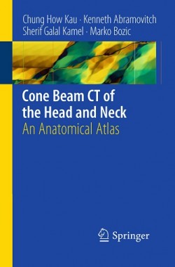
Cone Beam CT of the Head and Neck’ presents normal anatomy of the head using photographs of cadavers and CBCT images in sagittal, axial and coronal planes with the anatomic structures and landmarks clearly labelled. Important structures and regions are presented in detailed view. This is the first book with photographs of human tissue (based on slicing of cadaveric heads) combined with CBCT images.
Product Details
- Paperback: 75 pages
- Publisher: Springer; 1st Edition. edition (March 9, 2011)
- Language: English
- ISBN-10: 3642127037
- ISBN-13: 978-3642127038
- Product Dimensions: 5 x 0.3 x 7.4 inches
- Shipping Weight: 7.8 ounces
Download
Note: Only Dental member can download this ebook. Learn more here!


![[Free] Pocket Atlas of Sectional Anatomy, Computed Tomography and Magnetic Resonance Imaging, Vol. 1: Head and Neck, 3rd Edition](https://dental.downloadmedicalbook.com/wp-content/uploads/2012/06/Pocket-Atlas-of-Sectional-Anatomy-Computed-Tomography-and-Magnetic-Resonance-Imaging-Vol.-1-250x3741-150x150.jpg)

This is the book that helpful for everybody
friend can u please provide any book on cone beam ct physics and techniqe..thanx
I’ll try to find it!
Download site transfers trojan horse, please reupload to another provider.
New link added!