 By
By
- Robert P. Langlais, DDS, MS, FACD, FICD, Professor, Department of Dental Diagnostic Science, University of Texas Health Science Center at San Antonio, School of Dentistry, San Antonio, Texas
- Craig S. Miller, DMD, MS, FACD, Professor of Oral Medicine, Department of Oral Health Practice and Department of Microbiology, Immunology and Molecular Genetics, College of Dentistry, College of Medicine, University of Kentucky, Lexington, Kentucky
- Jill S. Nield-Gehrig, RDH, MA, Dean Emeritus, Division of Allied Health, Asheville-Buncombe Technical Community College
Asheville, North Carolina
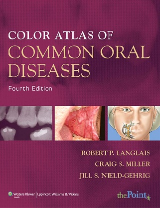
The Fourth Edition of the Color Atlas of Common Oral Diseases has been thoroughly revised, updated, and expanded with over 650 high-quality color photographs and radiographic illustrations of oral disease to help you recognize and identify oral diseases. It presents clinical and radiographic features of common diseases found in the oral cavity according to location, color, surface change, and radiographic appearance.
New to This Edition
- New! Learning objectives beginning each chapter focus your studies by setting forth what you’ll know upon successful completion of the chapter.
- New! Color-coded sections with tabs organize information into logical sections and help you locate materials quickly.
New! Highlighted key words draw your attention to important concepts. - New! Case studies at the end of each section ask you to apply your knowledge to resolving clinical situations and prepare you for the National Board Examinations.
- NEW! Greatly expanded Glossary offers clear, simple definitions for key terms.
- NEW! New topics are covered including malocclusion, temporomandibular joint disorders, drug-related oral lesions, and dental resorption.
- NEW! Ancillaries are available – Instructors have access to a test generator, an image bank, disease facts for selected chapters, answers to case studies, PowerPoint slides, and the full text online. Students also have access to the full text online. All resources are available at http://thePoint.lww.com/Langlais4e.
Key Features
- Exceptional value for its high quality and content.
- Over 650 high quality color photographs and radiographic illustrations carefully selected to represent features of conditions or diseases. High quality line drawings accompany these illustrations to help clarify the photos and radiographs.
- Presented in an easy-to-navigate format. Each color plate consists of 8 illustrations per page so disorders closely related by appearance can be easily compared.
- Concise overviews, on the page facing the illustrations, emphasize the clinical description (signs and symptoms) of oral lesions. They also discuss the nature of various disease processes and other clinically relevant data (sex, age, race affected by the disorder). Also included is a brief discussion of cause, including specific gene defects and pathophysiological abnormalities, and treatment methods. This helps to integrate oral diagnosis, medicine, pathology and radiology.
- Classifies diseases and conditions by the clinical appearance or by specific anatomic location.
Download
Note: Only Dental member can download this ebook. Learn more here!
Dental eBooks’ Library







Hello,
Well,Can i know whether dental journals like British dental journal,australian dental journals and like many others available to gold membership?
thank you.
Murali
Both are available but Australian dental journal takes too much time to download
Thank u