 By
By
Friedrich A. Pasler, DDS, PhD, Professor Emeritus, Department of Radiology, Dental Institute, University of Geneva, Switzerland;
Heiko Visser, Dr. med. dent., MSc Physics, Professor, Dental School, University of Göttingen, Germany
Book Description
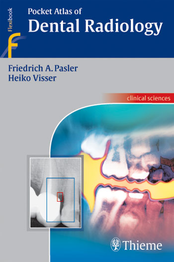
In this age of highly specialized medical imaging, an examination of the teeth and alveolar bone is almost unthinkable without the use of radiographs. This highly informative and easy-to-read book with a collection of 798 radiographs, tables, and photos provides a myriad of problem-solving tips concerning the fundamentals of radiographic techniques, quality assurance, image processing, radiographic anatomy, and radiographic diagnosis. Information is easy to find, enabling the reader to literally get a grasp of essential new knowledge in next to no time. The dental practice team now has a pocket “consultant” at its fingertips, providing practical ways to incorporate new techniques into daily practice.
Key Feature
A fine-tuned didactic concept
Each topical concept is printed compactly on a double-page spread
On the left: concise and highly instructive text
On the right: informative, high-quality illustrations
Main topics include:
Examination strategies, radiation protection, quality assurance
Conventional and digital radiographic techniques
Radiographic anatomy: The problems of object localization and how to solve them
Recent research with conventional radiography, CT, MRI, etc.
Normal variations and pathologic conditions as viewed with the various imaging techniques
A concise and up-to-date presentation of modern dental radiology
Download
Note: Only Dental member can download this ebook. Learn more here!
Related Books
 Oral Radiology: Principles and Interpretation, 6th Edition
 Oral Radiology: Principles and Interpretation, 5th Edition
 Three-Dimensional Cephalometry: A Color Atlas and Manual
 Pocket Atlas of Oral Diseases
 Essentials Of Dental Radiography and Radiology
 Periodontal Surgery: A Clinical Atlas
 Color Atlas of Dental Medicine, Aesthetic Dentistry
 Textbook and Color Atlas of Salivary Gland Pathology: Diagnosis and Management
 Color Atlas Of Temporomandibular Joint Surgery
 Atlas of the Oral and Maxillofacial Surgery Clinics of North America 2002-2004
link dead . please re upload it again
Updated
Hey Bear,
Link is broken, could u please reupload the file many thanx
New link updated!
Ryushare not working, I need this book, please update
Hi Bear can u reupload the link please?
New link added!