 By
By
Suresh K. Mukherji, M.D., Division Director of Neuroradiology and Head & Neck Radiology, Neuroradiology Fellowship Program Director, University of Michigan, Ann Arbor, Michigan
Vincent Chong, M.D., Program Director, DO Diagnostic Radiology, Singapore General Hospital, Singapore
Book Description
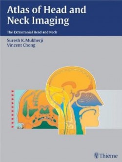
With hundreds of high quality illustrations, this book makes the identification and localization of complex neck masses relatively simple. This book provides CT and MR examples for more than 200 different diseases of the suprahyoid and infrahyoid neck, as well as clear and concise information on the epidemiology, clinical findings, pathology, and treatment guidelines for each disease.Each space within the head and neck has its own separate section, with examples of the common pathology that arises in this area. A standard format consisting of Epidemiology, Clinical Presentation, Pathology, Treatment, and Imaging Findings, allows quick and efficient access to well-structured subjects. This uniform organization streamlines research for radiologists at any level of training.Although well over 200 pathologies are included within this remarkable text, Atlas of Head and Neck Imaging focuses primarily on the suprahyoid and infrahyoid neck, providing exceptionally detailed information on the most challenging aspects of this field.
Editorial Reviews
Review
“..this text is an excellent textbook of extracranial head and neck imaging that could benefit mxillofacial radiologist.” — Journal of Maxillofacial Radiology, April 2005
“This book proved hard to fault…pursues the topic to an authoritative depth and is highly recommended.” — The Journal of Otolaryngology of Otology, April 2005
Review
Radiologists and radiation oncologists will find this visual text ideal as a quick anatomic reference and diagnostic tool. Radiology residents preparing for board exams and neuroradiology fellows and staff studying for the CAQ exam will also benefit from the wealth of information.
Customer Reviews
[…] Book Review May 21, 2010
This is a very useful text with a comprehensive review of radiologic findings in Head & Neck pathologic conditions. It is divided into the various neck spaces and each chapter within these sections deals with a specific pathologic finding for that area of the head and neck. The chapters are arranged from benign to malignant pathology and cover epidemiology, clinical findings, pathology, treatment, imaging findings and imaging pearls. The chapters are concise and the images are clear with detailed legends. The line drawings and artist renditions are easily followed and surgical or endoscopic views, where added, enhance the radiologic images presented.This text is useful for radiology residents, neuroradiology fellows, radiologists who perform head and neck imaging and otolaryngologists. It is a useful addendum to an otolaryngologist’s library and can enhance the review of patients’ films to determine the site and origin of a lesion. Radiation oncologists and medical oncologists who treat head and neck carcinomas will also find this text to be a useful resource. Oral maxillofacial surgeons would additionally benefit from the information in this book, especially with regards to the odontogenic infections and tumors. Because the chapters are clear and concise, the book is easy to read and can enhance other references to the specific pathology one is interested in.The authors specifically state in the preface that the purpose of this book is to be a reference used at the viewbox (or PACS machine) and can help identify the location with respect to the various neck spaces as well as the differential diagnosis. The inclusion of imaging pearls is something that residents especially will appreciate. I feel the authors accomplished their goal of making this a workable reference tool and their inclusion of CT, MRI as well as artist renderings and line drawings enhance the ability of this book to be used when reviewing patient images.Comparing this to other similar references, in specific Valvassori Imaging of the Head and Neck, I find the chapters easier to follow and the organization speeds up the search process for a specific pathology. Even though this book does not specifically address sinus or temporal bone imaging, I feel it is a useful adjunct to Valvassori’s text and for quick reference, it is clearly better.
Product Details
- Hardcover: 216 pages
- Publisher: Thieme; 1 edition (January 22, 2004)
- Language: English
- ISBN-10: 1588901785
- ISBN-13: 978-1588901781
- Product Dimensions: 12.2 x 9.3 x 1.3 inches
- Shipping Weight: 6.5 pounds
Download
Note: Only Dental member can download this ebook. Learn more here!

![[Free] Pocket Atlas of Sectional Anatomy, Computed Tomography and Magnetic Resonance Imaging, Vol. 1: Head and Neck, 3rd Edition](https://dental.downloadmedicalbook.com/wp-content/uploads/2012/06/Pocket-Atlas-of-Sectional-Anatomy-Computed-Tomography-and-Magnetic-Resonance-Imaging-Vol.-1-250x3741-150x150.jpg)



![[Free] Cone Beam CT of the Head and Neck: An Anatomical Atlas](https://dental.downloadmedicalbook.com/wp-content/uploads/2012/06/Cone-Beam-CT-of-the-Head-and-Neck-An-Anatomical-Atlas-250x3801-150x150.jpg)


![[Free] Imaging of the Head and Neck, 2nd Edition](https://dental.downloadmedicalbook.com/wp-content/uploads/2012/06/imaging-of-the-head-and-neck-150x150.jpg)
File not found.
New link added!